Quick Answer
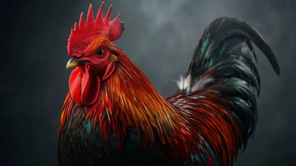
The viral internet phenomenon known as the “Zombie Chicken Breed” typically refers to the Ayam Cemani or Silkie, birds that owe their hyper-pigmented appearance to a genetic mutation called fibromelanosis. This condition involves the massive overexpression of the Endothelin 3 (EDN3) gene, which drives systemic hyper-migration of melanoblasts during embryonic development. This trait is entirely benign and hereditary, completely distinct from acute neurological pathogens that cause erratic motor function or ataxia.
Key Takeaways
- Fibromelanosis is driven by a complex genomic rearrangement involving a duplication upstream of the EDN3 locus.
- Melanoblast hyper-migration affects dermal, skeletal, and visceral tissues, turning them dark without impacting physiological viability.
- Internet myths conflating this genetic mutation with viral pathogens or infectious zombie states are biologically inaccurate.
- Standard breed registries maintain strict phenotypic criteria for fibromelanistic poultry that separate them from pathologically compromised birds.
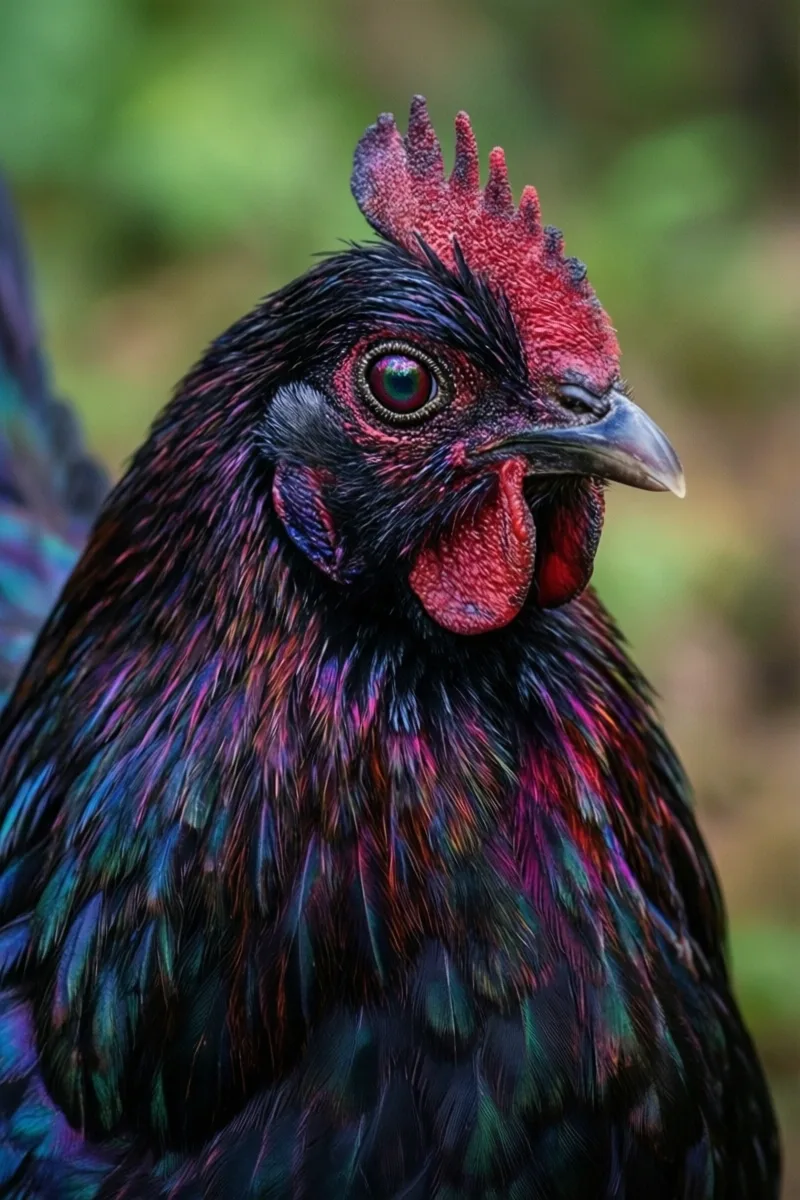
The colloquial internet designation of a “Zombie Chicken Breed” is a scientifically inaccurate moniker applied to rare, highly pigmented avian phenotypes like the Ayam Cemani, masking a fascinating and well-documented genetic reality. To understand why these birds exhibit an entirely black exterior and interior—spanning feathers, skin, periosteum, and internal organs—we must examine avian developmental biology rather than sensationalized folklore. Avian geneticists classify this unique presentation not as a pathological aberration, but as a stable, hereditary condition governed by specific molecular pathways that dictate cellular migration during embryogenesis.
Deconstructing the Fibromelanosisigration and EDN3 Overexpression
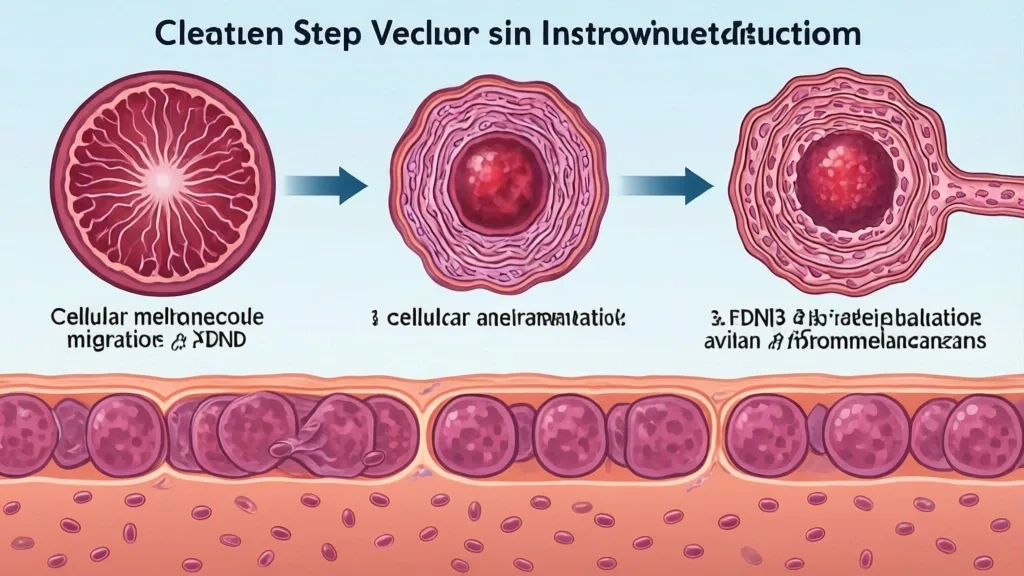
Fibromelanosis is a complex dermal hyperpigmentation trait controlled by a multi-gene complex, the key driver of which is the Endothelin 3 (EDN3) gene. In normal avian embryogenesis, neural crest cells give rise to melanoblasts—the precursors to pigment-producing melanocytes. These cells migrate along specific dorsolateral pathways into the epidermis. However, in fibromelanistic poultry, a massive genomic rearrangement occurs. Specifically, a complex mutation involving a duplication of approximately 140 kilobases on chromosome 20 places an additional regulatory sequence upstream of the EDN3 gene.
This structural mutation causes a dramatic, sustained upregulation of EDN3 transcription within the embryonic skin and surrounding connective tissues. Because EDN3 acts as a potent mitogen and survival factor for melanocyte precursors, its overexpression triggers a massive proliferation and hyper-migration of melanoblasts throughout the developing organism. Instead of restricting melanin deposition to standard epidermal structures like feathers and outer skin layers, the excess melanocytes infiltrate deep somatic tissues.
Consequently, the hyper-migration reaches the periosteum (the dense membrane covering bones), the walls of major blood vessels, the mesentery, and the capsule of visceral organs such as the liver and gizzard. Histological examination reveals heavily pigmented melanocytes densely packed within collagenous matrices and fascial planes. This biochemical pathway is robust, highly heritable, and completely distinct from acquired pathological pigmentation disorders or necrotic tissue changes often associated with infectious agents in poultry science.
Genotypic Reality versus Viral Internet Sensationalism
Social media platforms frequently propagate the myth that these hyper-pigmented birds represent an undead anomaly or a genetically engineered hybrid. Empirical avian genetics soundly refutes these claims. The trait is ancient, having been documented in Southeast Asian poultry populations for centuries before formal scientific isolation. Established agricultural bodies, including the Indonesian Livestock Directorate and the American Poultry Association, maintain strict standardizations for breeds like the Ayam Cemani and Silkie that outline these exact phenotypic parameters without invoking sensationalism.
From a metabolic and physiological perspective, fibromelanistic birds possess normal biological viability. Their oxygen transport, systemic immunity, and neurological processing function identically to standard wild-type or commercial poultry lines. The excess melanin deposition within visceral structures does not impede organ functionality, nor does it alter the nutritional composition of the meat or eggs. Backyard keepers and rare breed preservationists must differentiate between stable, centuries-old genetic mutations preserved through selective breeding and abnormal physiological states that require veterinary quarantine or therapeutic intervention.
Comparative Matrix: Fibromelanistic Traits vs. Pathological Neurological Syndromes
Viral content creators frequently blur the lines between normal genetic pigmentation and sick birds exhibiting neurological distress, falsely labeling both under the “zombie” umbrella. Scientifically, a stark division exists between hereditary melanistic presentation and acute neuropathology. The following matrix contrasts the biological markers of normal fibromelanosis against common avian pathogens that induce genuine behavioral and motor abnormalities.
| Biological Parameter | Hereditary Fibromelanosis (e.g., Ayam Cemani, Silkie) | Acute Neurological Pathology (e.g., Newcastle Disease, Marek’s) |
|---|---|---|
| Etiological Origin | Stable chromosomal mutation (EDN3 gene duplication). | Viral infection (Paramyxovirus, Herpesvirus) or neurotoxins. |
| Tissue Presentation | Systemic black pigmentation of dermis, periosteum, and viscera. | Normal tissue coloration; potential macroscopic lesions or tumors. |
| Behavioral Phenotype | Alert, active, normal motor coordination and foraging instincts. | Ataxia, torticollis (wry neck), tremors, paralysis, or lethargy. |
| Transmission Dynamics | Mendelian inheritance patterns governed by selective breeding. | Contagious horizontal transmission via aerosols, fomites, or dander. |
| Prognosis & Viability | Normal life expectancy and physiological health. | High mortality or permanent neurological deficits; often fatal. |
Evaluating poultry health through an empirical lens ensures that hobbyists and researchers do not misinterpret normal breed characteristics as clinical emergencies. By understanding the upstream regulatory mechanisms of EDN3 and the exact manifestations of avian neuropathologies, poultry keepers can maintain high standards of flock management anchored in biological fact rather than digital myths.
Diagnostic Procedures for Distinguishing Genetics from Neurological Pathology
Backyard hobbyists frequently confuse the systemic hyperpigmentation driven by fibromelanosis with acute neurological deterioration. While fibromelanotic breeds exhibit uniform dermal, periosteal, and visceral melanization governed by genetic transcription, acute behavioral aberrations indicate distinct neuropathology. Establishing a rigorous differential diagnosis requires systematic evaluation of locomotor function, postural anomalies, and systemic necropsy findings.
Step 1: Clinical Observation and Gait Analysis
Observe unprovoked locomotion in an open, slip-resistant enclosure. Document ataxia, torticollis, or unilateral paresis. Neurological vectors such as Marek’s disease virus or Avian Encephalomyelitis typically present as asymmetric limb paralysis or head tremors. Conversely, genetically hyperpigmented birds maintain symmetrical motor function, provided no secondary orthopedic or infectious compounding factors exist.
Step 2: External Integumentary and Ocular Examination
Inspect the sclera, iris, and pupillary light reflex. In Marek’s disease (neurolymphomatosis), ocular infiltration leads to irregular pupillary margins (“gray eye”) and blindness. In contrast, the Ayam Cemani and related fibromelanistic phenotypes maintain normal physiological ocular reflexes despite intense dark pigmentation of the iris, earlobes, and comb.
Step 3: Post-Mortem Gross Pathology and Histology
Perform a systematic necropsy to differentiate internal melanosis from pathological lesions. Fibromelanosis yields a uniform, slate-black or blue-black coloration across the visceral peritoneum, mesentery, and connective tissues without nodular disruption. Visceral lymphoid leukosis or Marek’s-induced visceral tumors present as discrete, grayish-white nodular or diffuse neoplastic masses on the liver, spleen, and kidneys, completely overriding normal tissue architecture.
Managing Flock Biosecurity and Genetic Lineage Preservation
Maintaining pure fibromelanistic lines while protecting a closed flock from epizootic outbreaks demands strict adherence to biosecurity protocols. Genetic integrity depends on managing the complex locus responsible for the Endothelin 3 (EDN3) duplication. Unregulated outcrossing rapidly dilutes the hyperpigmentation phenotype, reverting dermal and visceral melanin deposition to baseline wild-type levels within generations.
Flock managers must implement rigorous quarantine procedures for incoming stock. All external birds require a minimum thirty-day isolation period paired with diagnostic screening. Utilizing Polymerase Chain Reaction (PCR) testing helps detect subclinical viral vectors such as Avian Leukosis Virus (ALV) and Marek’s Disease Virus (MDV), which often mask genetic vigor with neuro-pathological morbidity. Furthermore, nutritional optimization is critical; severe deficiencies in Vitamin E, Thiamine (B1), or Selenium induce acute encephalomalacia and polyneuritis, producing clinical symptoms of neurological collapse that mimic viral pathogens.
Authoritative Reference Frameworks and Standards Boards
Rigorous poultry science relies on standardized nomenclature and peer-reviewed genomic datasets rather than viral internet hyperbole. Researchers and advanced breeders reference genetic libraries managed by major bioinformatics authorities to track structural variants and chromosomal translocations associated with avian pigmentation anomalies.
Key institutional frameworks guiding these evaluations include the National Center for Biotechnology Information (NCBI) genomic databases, which house sequencing data for the FM locus and EDN3 overexpression models. Additionally, phenotypic validation standards maintained by avian pathology manuals and poultry breeders’ federations provide the baseline parameters required to assess true breed characteristics separate from infectious disease states.
📌 Save to Pinterest
Frequently Asked Questions
What genetic mutation causes the black meat and bones in the Ayam Cemani?
The phenotype is caused by fibromelanosis, a complex genetic trait involving a massive duplication of the Endothelin 3 (EDN3) gene. This duplication drives a hyper-proliferation and widespread migration of melanoblast precursors from the neural crest into embryonic tissues where melanin is not typically expressed, including periosteum, internal organ membranes, and connective tissue.
Are chickens labeled as zombie breeds actually suffering from neurological disease?
No. Sensationalized internet labels confuse normal genetic traits with pathology. Breeds like the Ayam Cemani exhibit completely healthy motor function and normal physiological behavior despite their striking external and internal black pigmentation, which is an inherited developmental trait rather than a sign of neurological decay.
How does Endothelin 3 (EDN3) regulate melanocyte migration in poultry?
Endothelin 3 acts as a potent paracrine growth factor for melanocyte precursors. In fibromelanistic poultry, regulatory mutations lead to a dramatic upregulation of EDN3 transcription in the dermis and connective tissues, signaling neural crest-derived melanoblasts to proliferate excessively and migrate throughout the entire organism during embryogenesis.
What is the structural difference between fibromelanosis and normal dermal melanin?
Normal dermal melanin is typically restricted to the epidermis, feathers, and specific localized skin patches under direct hormonal or genetic control. Fibromelanosis, by contrast, is a systemic developmental condition that penetrates deep connective tissue layers, encasing the walls of major blood vessels, nerves, and visceral organs in dense layers of true melanin pigment.
Can standard backyard biosecurity prevent acute neurological pathologies mistaken for genetic traits?
Strict biosecurity—including all-in-all-out flock management, rigorous quarantine protocols, vector control, and sanitized brooding environments—prevents infectious neurological agents like Marek’s disease virus and Avian Encephalomyelitis. However, biosecurity does not alter or prevent genetic conditions, as fibromelanosis is fixed at the chromosomal level during embryonic development.